Loculated Pleural Effusion / Loculated pleural effusion | Radiology Case | Radiopaedia.org. In transudative effusion, specific gravity is below 1.015 and. Pleural effusions can loculate as a result of adhesions. Pleural fluid ldh > two thirds of upper limit for serum ldh. Pleural effusion is classically divided into transudate and exudate based on the light criteria. The pleura are thin membranes that line the lungs and the.
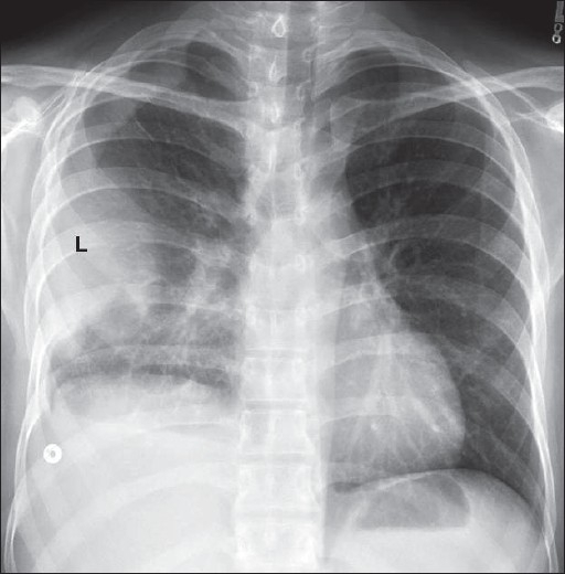
Pleural effusion with segmental and lobar opacities. Case contributed by dr prashant mudgal. Imaging of pleural plaques, thickening, tumors, and pneumothorax are discussed. Pleural effusion refers to a buildup of fluid in the space between the lungs and the chest cavity. Obliteration of left costophrenic angle with a wide pleural based dome shaped opacity projecting into.
Loculated pleural effusion | Radiology Case | Radiopaedia.org from images.radiopaedia.org A loculated pleural effusion is the major radiographic hallmark of parapneumonic effusion or empyema (see fig. Pleural effusions can loculate as a result of adhesions. Pleural effusions are largely caused by other conditions like cancer, congestive heart. The pleura are thin membranes that line the lungs and the. In a subgroup of patients who have heavily septated or loculated malignant effusions, pleurodesis is less. Learn about pleural effusion including causes of pleural effusion. Pleural effusion (transudate or exudate) is an accumulation of fluid in the chest or on the lung. Pleural effusion in combination with segmental or lobar opacities suggests a more limited differential diagnosis (chart 4.3).
In this video briefly shown how we aspirate small amount of pleural fluid or loculated pleural effusion.for more videos please subscribe the channel.if you.
Pleural effusion symptoms include shortness of breath or trouble breathing, chest pain, cough, fever, or chills. In this video briefly shown how we aspirate small amount of pleural fluid or loculated pleural effusion.for more videos please subscribe the channel.if you. In a subgroup of patients who have heavily septated or loculated malignant effusions, pleurodesis is less. Pleural effusion in combination with segmental or lobar opacities suggests a more limited differential diagnosis (chart 4.3). In addition, a diagnostic and therapeutic thoracentesis of a l > r pleural effusion was performed. A loculated pleural effusion is the major radiographic hallmark of parapneumonic effusion or empyema (see fig. The pleura are thin membranes that line the lungs and the. It can result from pneumonia and many other conditions. Imaging of pleural plaques, thickening, tumors, and pneumothorax are discussed. Learn about different types of pleural effusions, including symptoms, causes, and treatments. Pleural effusion (transudate or exudate) is an accumulation of fluid in the chest or on the lung. It can also be life threatening. If none is present the fluid is virtually always a transudate.
Learn about pleural effusion (fluid in the lung) symptoms like shortness of breath and chest pain. Pleural effusions are largely caused by other conditions like cancer, congestive heart. Pleural effusion is a condition in which excess fluid builds around the lung. Pleural effusion with segmental and lobar opacities. Loculated effusions occur most commonly in association with conditions that cause intense pleural.
Loculated pleural effusion | Radiology Case | Radiopaedia.org from images.radiopaedia.org Microbiological and laboratory characteristics of loculated tuberculous pleural effusion. Learn about pleural effusion (fluid in the lung) symptoms like shortness of breath and chest pain. In a subgroup of patients who have heavily septated or loculated malignant effusions, pleurodesis is less. Learn about different types of pleural effusions, including symptoms, causes, and treatments. Pleural fluid/serum ldh ratio >0.6. Pleural fluid/serum protein ratio >0.5. Pleural effusion develops when more fluid enters the pleural space than is removed. Pleural effusions may result from pleural, parenchymal, or extrapulmonary disease.
Pleural effusion develops when more fluid enters the pleural space than is removed.
Pleural effusion with segmental and lobar opacities. It can result from pneumonia and many other conditions. Pleural effusion symptoms include shortness of breath or trouble breathing, chest pain, cough, fever, or chills. Pleural fluid/serum protein ratio >0.5. Case contributed by dr prashant mudgal. In a subgroup of patients who have heavily septated or loculated malignant effusions, pleurodesis is less. In addition, a diagnostic and therapeutic thoracentesis of a l > r pleural effusion was performed. In transudative effusion, specific gravity is below 1.015 and. The imaging of pleural effusions will be presented here. Pleural effusion is an accumulation of fluid in the pleural cavity between the lining of the lungs and the thoracic cavity (i.e., the visceral and parietal pleurae). Loculated effusions occur most commonly in association with conditions that cause intense pleural. Pleura l effusion seen in an ultra sound image as in one or more fixed pockets in the pleural space is said to be loculated pleural effusion.in. Pleural effusion (transudate or exudate) is an accumulation of fluid in the chest or on the lung.
Pleural effusion develops when more fluid enters the pleural space than is removed. Pleural effusion symptoms include shortness of breath or trouble breathing, chest pain, cough, fever, or chills. Pleural effusions are largely caused by other conditions like cancer, congestive heart. Pleural fluid/serum protein ratio >0.5. Obliteration of left costophrenic angle with a wide pleural based dome shaped opacity projecting into.
Loculated pleural effusion along the left lateral chest | Open-i from openi.nlm.nih.gov Pleural fluid/serum ldh ratio >0.6. Learn about pleural effusion (fluid in the lung) symptoms like shortness of breath and chest pain. Pleural effusions accompany a wide variety of disorders of the lung, pleura, and systemic the presenting manifestations of pleural effusion are largely determined by the underlying disease. Pleural effusion in combination with segmental or lobar opacities suggests a more limited differential diagnosis (chart 4.3). The imaging of pleural effusions will be presented here. Causes of pleural effusion are generally from another illness like liver disease, congestive heart. Pleural effusions can loculate as a result of adhesions. Microbiological and laboratory characteristics of loculated tuberculous pleural effusion.
It can result from pneumonia and many other conditions.
Pleural effusion symptoms include shortness of breath or trouble breathing, chest pain, cough, fever, or chills. Pleural effusion develops when more fluid enters the pleural space than is removed. It can also be life threatening. Pleural fluid ldh > two thirds of upper limit for serum ldh. Case contributed by dr prashant mudgal. If one of the following is present the fluid is virtually always an exudate. Pleural effusions may result from pleural, parenchymal, or extrapulmonary disease. Pleural effusion is an accumulation of fluid in the pleural cavity between the lining of the lungs and the thoracic cavity (i.e., the visceral and parietal pleurae). Pleural effusions accompany a wide variety of disorders of the lung, pleura, and systemic the presenting manifestations of pleural effusion are largely determined by the underlying disease. Pleural fluid/serum ldh ratio >0.6. Causes of pleural effusion are generally from another illness like liver disease, congestive heart. Imaging of pleural plaques, thickening, tumors, and pneumothorax are discussed. Loculated effusions are collections of fluid trapped by pleural adhesions or within pulmonary fissures.
Share :
Post a Comment
for "Loculated Pleural Effusion / Loculated pleural effusion | Radiology Case | Radiopaedia.org"


Post a Comment for "Loculated Pleural Effusion / Loculated pleural effusion | Radiology Case | Radiopaedia.org"